[category]
[title]
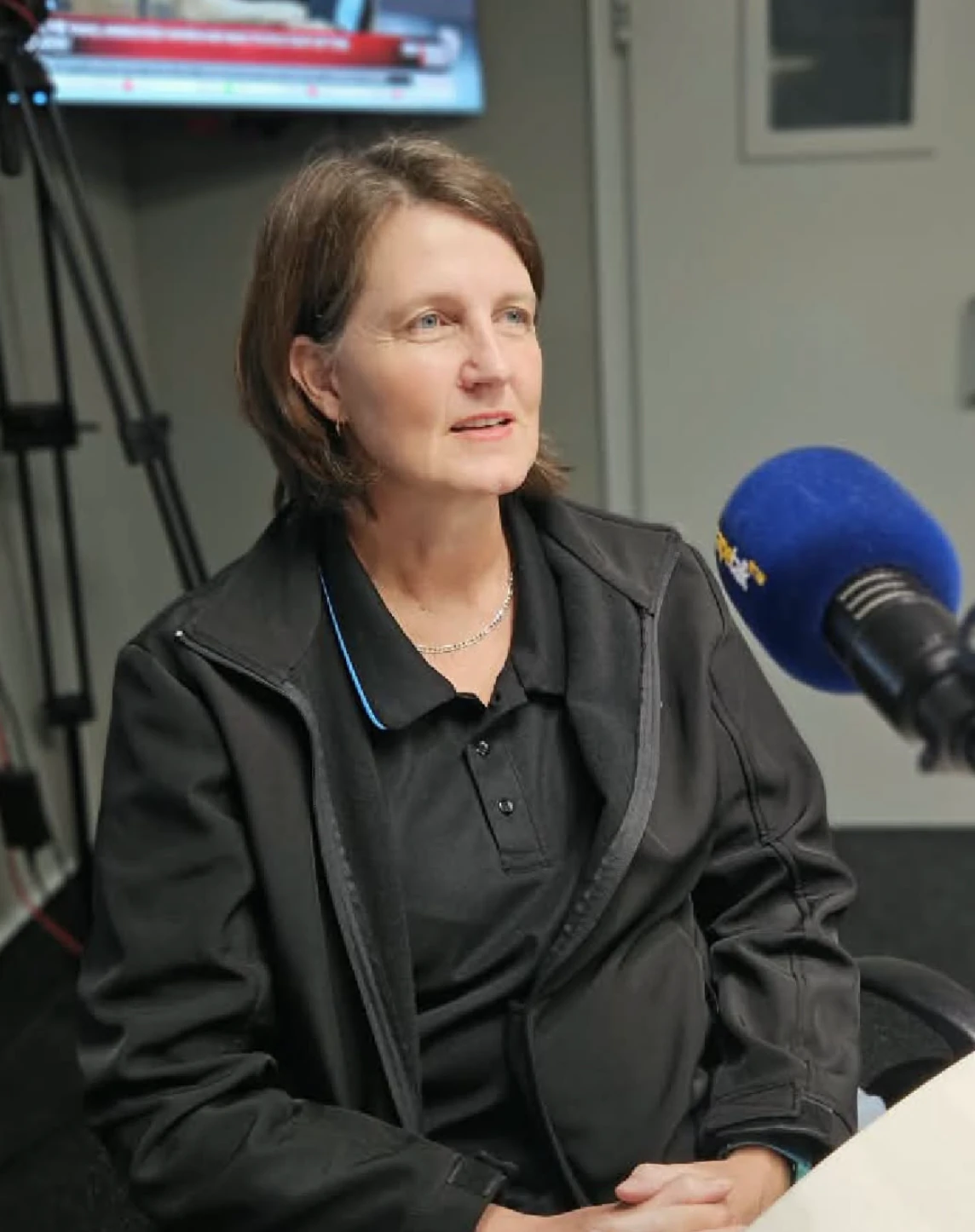
Dr Marna du Toit, whose European qualification brings international standards to South Africa’s breast cancer research, unpacks how dense breast tissue can delay diagnosis.


Seven months ago, South African radiologist Dr Marna du Toit graduated with the European Diploma in Breast Imaging (EDBI) from the prestigious European Board of Radiology (EBR). Most of her work focuses on dense breast tissue and how it often makes it harder for healthcare professionals to detect breast cancer.
For Africa, Dr du Toit’s newly obtained diploma means that the breast cancer tracking data, biopsy records and screening results from clinics in this country are internationally compliant.
Describing dense breast tissue to Time Out, the doctor said this means that a woman has a higher amount of tissue in the glands and fibres. This is a normal occurrence affecting roughly 50% of women above the age of 40.
“Breasts are composed of fibrous tissue, glandular tissue and fatty tissue. If you are told that you have dense breasts, it means that you have more fibrous and glandular tissue and less fatty tissue,” the doctor explained.
In a mammogram, dense breast tissue shows up as solid white, as do breast tumours and abnormalities.
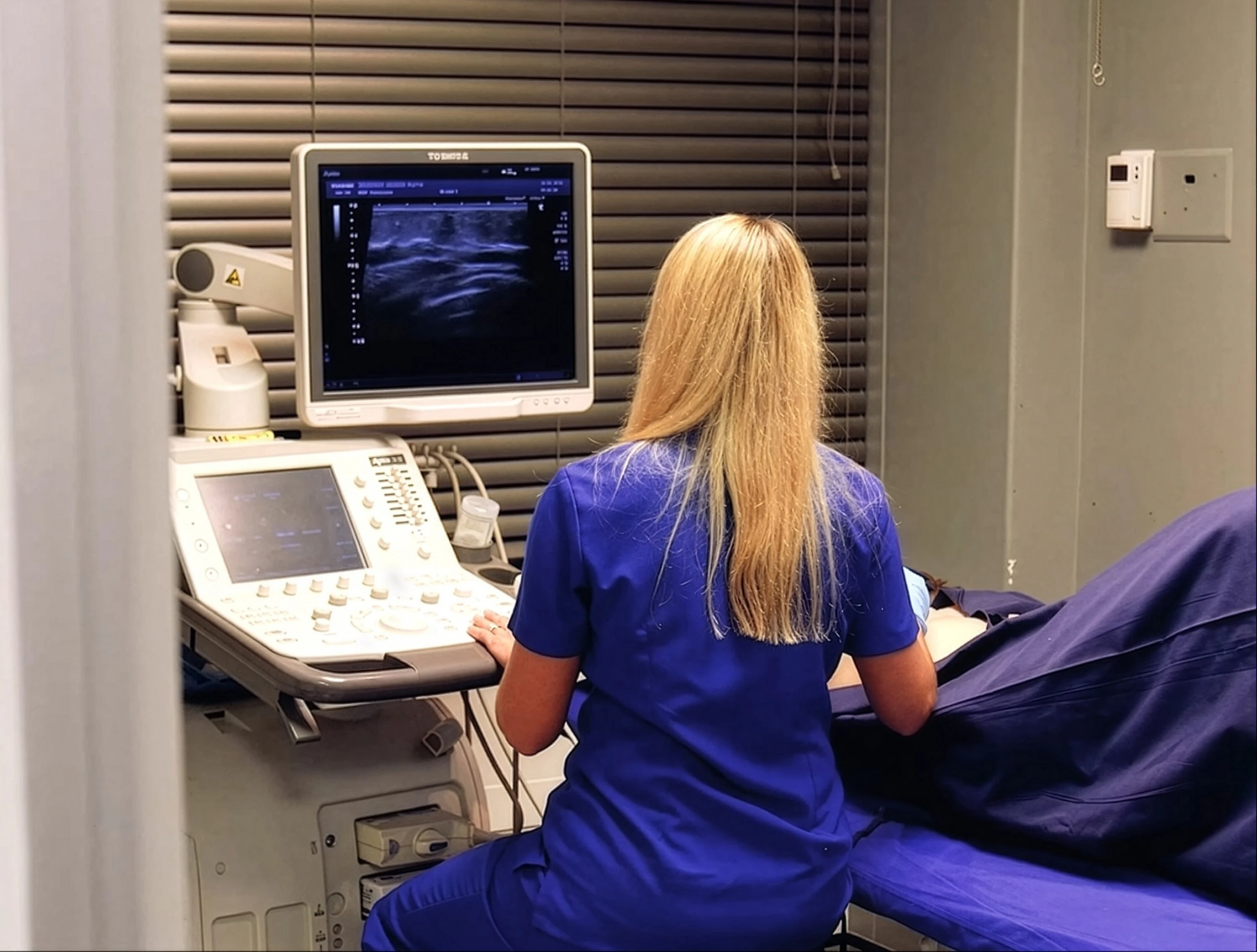
It is therefore recommended that women with dense breasts should not only get a standard mammogram, but should also supplement this examination with breast ulcersounds, 3D mammograms (tomosynthesis) or breast MRIs.
“High breast density is more common in younger women. However, the relative risk multiplier associated with dense tissue persists across all ages, though the proportion of women who retain high density decreases as they age,” she elaborated.
Studies have also shown that high breast density remains a risk factor in older and postmenopausal women.
Explaining the role of breast reduction in cancer detection, Dr du Toit said since this surgery reduces the overall volume of dense tissue in the breasts, having less tissue can lower women’s lifetime risk of developing breast cancer.
“While a breast reduction removes volume, the remaining tissue may still require alternative or supplemental screening methods (such as 3D mammograms, ultrasound or breast MRI) because the underlying density of the remaining tissue does not change.”
The doctor disputed claims that have been made by various individuals on social media about breasts growing back after reduction, saying the factors that contribute to breasts getting larger after the surgery include weight gain, pregnancy or hormonal changes.
Dense tissue making abnormalities harder to detect can delay breast cancer diagnosis, but research shows dense breasts also carry an independent biological risk. However, breast density alone does not predict the risk of breast cancer; it is just one piece of the puzzle.
Follow Time Out Johannesburg on Facebook, TikTok and Instagram! While you're at it, sign up for our newsletter to receive even more of the best of your city.
TIME OUT JOHANNESBURG RECOMMENDS:
📍 The best things to do in Johannesburg
🏨 The best hotels in Johannesburg
🍽 The best restaurants in Johannesburg
🛏️ Where to stay in Johannesburg
📢 Latest news from Johannesburg
Discover Time Out original video